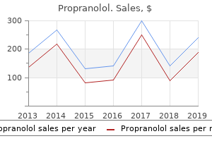
"Purchase propranolol, cardiovascular system video animation".
By: Z. Temmy, M.A., M.D., M.P.H.
Vice Chair, Weill Cornell Medical College
Step 2 cardiovascular disease genetic 20mg propranolol free shipping, Oxidation of Iodine the iodide taken up by the thyroid cell is oxidised to active iodine (Step 2 in arteries by stina propranolol 40 mg fast delivery. In patients with an inborn error of iodide oxidation defect jaw arteries discount propranolol online visa, treatment is T4 administration blood vessels of the eye propranolol 40 mg sale. There are 115 tyrosine residues in the Tgb, out of which 35 residues can be iodinated. Iodination of the tyrosine is taking place on the intact Tgb molecule in the follicular space. Step 4, Coupling Some of the tyrosine residues in the thyroglobulin are aligned opposite each other, and are coupled (step 4. The iodination and coupling are taking place in the borders of the follicular cells. Step 5, Storage the thyroid gland is unique, in that it is the only endocrine gland to store appreciable amounts of the hormone (Step 5 in. Step 6, Utilization When necessity arises, the thyroglobulin is taken from the acinar colloid, into the cell by pinocytosis (Step 6. This hydrolysis is depressed by iodide and therefore potassium iodide (Kl) is used as an adjuvant in hyperthyroidism. In a genetic disorder, abnormal Tgb is synthesized, resulting in deficient proteolysis and deficiency of thyroxine. Since 540 Textbook of Biochemistry; Section F: Hormones iodine is excreted, iodine deficiency is manifested. Step 10, Transport of Thyroid Hormones Thyroid hormones are transported in plasma by proteins (Step 10 in. Step 11, Catabolism of Thyroid Hormones T4 has a half-life of 4-7 days, while T3 has about 1 day. Part of the T3 and T4 are conjugated with glucuronic acid and excreted through bile, and to a lesser extent, through urine. Deamination of T4 produces tetraiodothyroacetic acid (Tetrac); and T3 gives rise to tri-iodo-thyroacetic acid (Triac). Higher concentration of T3 causes protein catabolism and negative nitrogen balance. Cholesterol degradation is increased and hence cholesterol level in blood is decreased, which is another hallmark of hyperthyroidism. Mechanism of Action of Thyroid Hormone the hormone attaches to specific nuclear receptors. Advanced techniques are chemiluminiscence and immunofluorescence; these also utilize antibodies for the assay technique. The values of free hormones are not affected by the amount of carrier proteins in the blood. Binding proteins Availability of the assay of free hormone has made this test only of historical importance. The abnormalities in the level of binding proteins may be reflected as abnormal hormone increase increase decrease decrease marked normal increase marked normal decrease normal normal increase decrease Pregnancy increase decrease decrease increase normal increase mild normal increase Chapter 47; Thyroid Hormones 541 Table 47. Since sensitive and accurate assay techniques are available now for measurement of free T4 and free T3, the fallacies due to alteration of binding proteins do not affect the assessment of the functional status of the gland. It is not diagnostic, because hypercholesterolemia is seen not only in hypothyroidism, but also in diabetes mellitus, hypertension, obstructive jaundice and nephrotic syndrome. However, cholesterol level is a useful index in monitoring the effectiveness of the therapy in thyroid conditions. Cholesterol level is increased in hypothyroidism, because cholesterol carrying lipoprotein degradation is decreased. If the hypothalamopituitary-thyroid axis is normal, the T3 and T4 secretions will be increased. An exaggerated response is observed in primary hypothyroidism since the negative feedback effect of T4 is reduced. Abnormalities of Thyroid Function In 1835 Robert James Graves and in 1840 Carl Adolph Basedow described the hyperthyroidism (Graves-Basedow disease). Emil Kocher was the first surgeon to excise thyroid gland to treat goiter in 1883. Diseases of the thyroid are the most common afflictions involving the endocrine systems. The commonest types of thyroid diseases are hyperthyroidism (excess secretion), hypothyroidism (decreased secretion) and goiter (enlargement of thyroid gland).
On the other hand cardiovascular disease what causes it order propranolol pills in toronto, two unrelated individuals would possess virtually no markers in common cardiovascular system jeopardy ppt discount 20mg propranolol visa. Scattered through the human genome are stretches containing repeats of a 32 base pair sequence capillaries 3 types order propranolol 80mg free shipping. In any one of the stretches the sequence is repeated over and over again cardiovascular system nursing interventions buy propranolol 80 mg low price, sometimes reaching a length of thousands of nucleotides. Different individuals possess these repeated sequences, called minisatellites, in the same locations, but the number of repeats of the short sequence varies from person to person. The fragments containing the repeated sequence are then identified by Southern transfer using a radioactive probe containing the 32 base repeated sequence. For a typical individual, the cleaving and probing procedure resolves about 20 fragments of size greater than several thousand base pairs. Since each individual possesses different numbers of the 32 base repeats in these long stretches, each possesses a different collection of sizes for these large fragments. Megabase Sequencing In addition to mapping, sequencing often provides fundamental information for further studies on a gene or gene system. The sequencing techniques described in the previous chapter are adequate for sequencing a few genes, but one area of great interest, the immune system, possesses hundreds of genes, many of unknown function. Serious effort is now also going into the early steps of determining the sequence of the entire human genome. Large sequencing projects such as these require better methods, and several have been developed. The one described below eliminates the use of radioisotopes and automates the detection of the bands on gels. These are obtaining the plasmids necessary for sequencing the desired region, pouring the gels, exposing and developing the autoradiograph films, and reading the information from the films. Imagine the savings in the Sanger sequencing technique if each of the four dideoxynucleotides could be tagged with a unique label. Then, instead of labeling the primer or the first nucleotides synthesized, the chain terminating nucleotide would possess the label. If this were done and each of the four labels were distinguishable, the four dideoxynucleotides could be combined in the same synthesis tube and the complex mixture of the four families of oligonucleotides could be subjected to electrophoresis in the same lane of the gel. Following electrophoresis, the four families of oligonucleotides could be distinguished and the entire sequence read just as though each one occupied a unique lane on the gel. Each of the four fluorescent groups that emits at wavelengths 1, 2, 3, and 4, would be attached to a different base. Excitation Emission 1 2 3 4 Wavelength 312 Advanced Genetic Engineering Electrophoresis Photomultiplier detector Glass plate Gel Laser excitation Figure 10. Furthermore, each of the four nucleotides must be modified with a different adduct, one that fluoresces at a different wavelength from the others. In addition, it is useful if the excitation spectrum of the four fluorescent molecules substantially overlap so that only one exciting wavelength is required. Although the entire gel could be illuminated following electrophoresis, it is easier to monitor the passage during electrophoresis of one band after another past a point near the bottom of the gel. By measuring the color of the fluorescence passing a point near the bottom of the gel, the nucleotide terminating this particular size of oligonucleotide can be determined. Multiple lanes can be monitored simultaneously so that the sequence can be determined semiautomatically of many different samples simultaneously. A more serious problem than the sensitivity is the generation of useful samples to be sequenced. This shotgun approach yields the desired sequences if sufficient clones are available and sufficient time and effort are expended. When a few gaps remain, it may be easier to close them by chromosome walking than by sequencing more and more Footprinting, Premodification and Missing Contact Probing 313 Cloned fragment Sequencing primer Cut again and reclone the shortened fragment Sequencing vector Cut with restriction enzyme and digest with exonuclease Figure 10. The vector is opened and digested, then a fragment is removed by cutting a second time with a restriction enzyme. This fragment is recloned and sequence is determined by using a primer to a sequence within the cloning vector adjacent to the location of the inserted fragment. By performing a series of exonuclease digestions for increasing periods, progressively larger deletions may be obtained. Another method of generating the necessary clones for sequencing a large region is to use a nested set of overlapping deletions. By sequencing from a site within the vector sequences with the use of an oligonu cleotide that hybridizes to the vector, the first 400 or so nucleotides of each of the clones can be determined.
Such signals might be as simple as cells keeping track of their ancestors cardiovascular system ventricles generic propranolol 20 mg with amex, or as complicated as schemes involving signaling between groups of cells cardiovascular system failure discount 40 mg propranolol mastercard. The simplest form of general signaling would seem to be to use chemicals cardiovascular exercise program 80mg propranolol with mastercard, which we shall call morphogens cardiovascular system while running cheap propranolol 80 mg with visa, whose concentrations can indicate positions. This would be much like specifying locations on the earth with latitude and longitude. General Considerations on Signaling In principle, three chemicals whose concentrations varied in the x, y, and z directions would be sufficient to specify every important location in a developing organism. After determining its position, any cell could induce or repress the genes appropriate to its position. Generating a simple coordinate system in which the locations of points are determined by the concentrations of three chemicals presents several problems. The embryo cannot leave it to chance that an appropriate cell will start off the process of building a gradient, perhaps by synthesizing and secreting some compound. Therefore, either certain cells are special as a result of their lineage and they will set up the gradients, or an external influence directs some of the cells in the embryo to behave differently from the rest. In either case, the embryo 479 480 Genes Regulating Development z y x Concentration Concentration y x Concentration z Figure 17. Commonly, at least one external influence or definition of an axis of the developmental coordinate system comes from the mother. They cannot be introduced into the egg during its development if the signal is a freely diffusing molecule. Any gradient in such a molecule would diffuse away before fertilization and embryo development. Therefore maternal effects either must be generated during development or they must be placed in the egg in a way that diffusion cannot alter. To reach all cells, the chemicals used to specify location in an embryo must be freely diffusible. If the gradients are set up in the egg after it has been subdivided into cells, the chemicals must enter and leave cells freely. On the other hand, position might be determined before a fertilized egg has divided into cells. Drosophila operates this way, and the embryo reaches about 4,000 nuclei before cell walls are synthesized. If many different coordinate positions are to be distinguished along one concentration gradient, precise measurements of the morphogen concentration must be made. The standard biochemical means for measuring concentration are simply measuring the amount of binding of a chemical to a receptor with appropriate affinity. This method is incapable of discerning small differences in the concentration of a substance, but detecting such differences would be necessary if many different developmental cues are to be derived from one gradient. Instead of using a few gradients to determine everything about a General Considerations on Signaling 481 Amount bound Bound = B2 B1 Concentration Kd + Concentration C1 C2 Concentration Figure 17. These can be subdivided as many times as necessary to produce as many different states as are required for the developing embryo. Not only must cells determine where they are in the coordinate system of morphogens, but they must do so crisply. Therefore we can expect developing organisms to use special techniques to make the division lines sharp. One simple technique is to make decisions when there are as few cells as possible. Another is to use a small embryo, for the smaller the embryo, the steeper the gradients, and therefore the easier it is to make decisions. Once an individual cell has determined what tissue or body part it is to become, it can go through multiple cell divisions to generate as much tissue as necessary. A second general way to make sharp dividing lines in differentiating tissue is to make the processes nonlinear. Suppose the decision between becoming tissue of type A or type B depends on the amount of morphogen bound to a protein, and that the binding is described by the Figure 17. Further, assume that all cells whose receptors bind less than x1 of the morphogen become cell-type A, and all cells whose receptors bind more than x2 of the morphogen become type B. They could use two receptors and respond to two gradients or respond to a single morphogen by requiring that two binding events occur.
The pH within red cells is lower than that of the surrounding plasma is due arteries 2012 buy discount propranolol 80mg on-line, in part karan capillaries pvt ltd faridabad discount propranolol on line, to the very high concentration of negative non-diffusible hemoglobin ions cardiovascular output purchase cheap propranolol. This will cause unequal distribution of H+ ions with a higher concentration within the cell arteries body buy cheap propranolol on line. When the cell membrane is disrupted, either by mechanical means or by lysing the membrane by Tween-20 (a lipid solvent), the organized particles inside the cell are homogenised. The organelles could then be separated by applying differential centrifugal forces (Table 2. Marker Enzymes Some enzymes are present in certain organelles only; such specific enzymes are called as marker enzymes (Table 2. After centrifugation, the separated organelles are identified by detection of marker enzymes in the sample. Nucleus is surrounded by two membranes: the inner one is called perinuclear membrane with. Typical cell 1= Nuclear membrane; 2= Nuclear pore; 3= Nucleolus; 4= endoplasmic reticulum; 5= Golgi body; 6= Mitochondria; 7= Microtubule; 8= Lysosome; 9= Vacuole; 10= Plasma membrane. Nucleus 8 Textbook of Biochemistry; Section A: Chemical Basis of Life numerous pores. In some cells, a portion of the nucleus may be seen as lighter shaded area; this is called nucleolus. It is a network of interconnecting membranes enclosing channels or cisternae, that are continuous from outer nuclear envelope to outer plasma membrane. Under electron microscope, the reticular arrangements will have railway track appearance. Microsomal cytochrome P-450 hydroxylates drugs such as benzpyrine, aminopyrine, aniline, morphine, phenobarbitone, etc. The rough appearance is due to ribosomes attached to cytoplasmic side of membrane where the proteins are being synthesized. Newly synthesized proteins are sorted first according to the sorting signals available in the proteins. Then they are packed into transport vesicles having different types of coat proteins. Finally, they are transported into various destinations; this is an energy dependent process. They are formed into a secretory vesicle, where these products are stored for a longer time. Release of trypsinogen by pancreatic Chapter 2; Subcellular Organelles and Cell Membranes 9 Table 2. Catalase and peroxidase are the enzymes present in peroxisomes which will destroy the unwanted peroxides and other free radicals. These crystals when phagocytosed, cause physical damage to lysosomes and release of enzymes. Following cell death, the lysosomes rupture releasing the hydrolytic enzymes which bring about postmortem autolysis. Cathepsins are normally restricted to the interior of lysosomes, but certain cancer cells liberate the cathepsins out of the cells. Then cathepsins degrade the basal lamina by hydrolysing collagen and elastin, so that other tumor cells can travel out to form distant metastasis. There are a few genetic diseases, where lysosomal enzymes are deficient or absent. Silicosis results from inhalation of silica particles into the lungs which are taken up by phagocytes. This stimulates fibroblast to proliferate and deposit collagen fibers, resulting in fibrosis and decreased lungs elasticity. Inclusion cell (I- cell) disease is a rare condition in which lysosomes lack in enzymes, but they are seen in blood. This means that the enzymes are synthesized, but are not able to reach the correct site.
However capillaries on face order propranolol 40mg visa, most adenocarcinomas are composed of more than one subtype cardiovascular system careers generic propranolol 20mg without a prescription, and the tumours 302 Chapter 5 coronary heart 6 guest purchase genuine propranolol on-line. The adenocarcinoma in situ subtype is characterized by lepidic (scale-like) growth along existing alveolar walls without underlying tissue invasion 10 cardiovascular risk propranolol 20 mg on line. The papillary subtype has fibrovascular cores, which distinguish it from the micropapillary subtype. No other molecular features have been described that separate these two common subtypes. Squamous cell carcinoma Squamous cell carcinoma has three subtypes: keratinizing, non-keratinizing, and basaloid. The morphological difference between the keratinizing subtype and the non-keratinizing Epigenetics of lung cancer the epigenetic landscape of lung cancer commences early during pathogenesis and consists of two major components: methylation and. Mutation spectra by histological type of lung cancer, showing the percentage of samples with a mutation detected by automated analysis. Several hundred genes are methylated in lung cancers, and the best studied and most frequently methylated genes are listed in Table 5. Methylation results in inactivation of one allele, and the other allele is usually deleted. In addition to methylation, many covalent modifications can occur on the N-terminal tail that protrudes from each of the four histone proteins. The genetic and epigenetic somatic alterations of lung cancer have recently been reviewed [37]. Morphological features of adenocarcinoma subtypes: (A) adenocarcinoma in situ, (B) acinar, (C) solid with mucin, (D) papillary, (E) micropapillary, and (F) mucinous. For example, adenocarcinomas are more prevalent in never-smoker patients with lung cancer [38]. In addition, lung cancers in never-smokers have different somatic characteristics. Overall, there are extensive differences between smokers and neversmokers with regard to the tumour 304 Chapter 5. Other features that distinguish lung cancer in never-smokers and eversmokers, such as methylation patterns, have also been reported [39]. However, in populations where the prevalence of smoking is low, an increasing proportion of lung cancer occurs in never-smokers and former smokers. This presented an appealing complementary strategy for reducing lung cancer mortality through detection of early-stage lung cancer, which is still potentially curable by surgical resection [40]. However, studies have shown that applying individual risk probability-based screening criteria could prevent more lung cancer deaths and reduce the number needed to screen to prevent one lung cancer death [42]. Although substantial efforts have been made to establish lung cancer risk prediction models based on personal health and exposure history [43], lung cancer researchers are now working towards integrating individual molecular profiles to improve risk prediction. Although most of the biomarkers have failed to be replicated in independent studies, several promising biomarkers have been established across multiple prospective cohort studies. For example, plasma level of pro-surfactant protein B was shown to be an independent predictor of lung cancer risk based on a pan-Canadian screening programme and the Carotene and Retinol Efficacy Trial, after adjusting for demographic factors and lung cancer risk factors [44]. It has become clear that a panel of multiple biomarkers, rather than any single marker, would be needed to improve risk prediction [45]. A succinct review of various reported biomarker panels was recently published [46]. In addition to blood-based biomarkers, another type of biomarker for early detection of lung cancer focuses on the gene expression profile of the airway epithelium, based on the theory of field of injury and field cancerization [48,49]. Finally, given the known association between chronic obstructive pulmonary disease and risk of lung cancer, previous studies have evaluated the added predictive performance of lung function [50,51]. It is anticipated that biomarkers may also help to differentiate malignant nodules from benign ones. The challenge is to establish a panel that would be applicable in the clinical setting and remain cost-effective for the health-care system. Information Commons Exposome Genome Informed mechanistic studies Integration Transcriptome Epigenome Microbiome Metabolome Clinical information Epidemiological data Biocomputing Observational clinical studies Knowledge Network Target identification New molecular taxonomic classification of patients by biomarkers Guide Guide Improve Diagnosis Treatment Diagnosis Molecular mechanisms Health outcomes Biomedical research Prevention research Clinical medicine 306 Chapter 5. In response to the need to differentiate between benign and malignant nodules, radiomics has emerged as a field of study.
Generic propranolol 80 mg on-line. Cardiac Exam - Point of Maximal Impulse.